Showing posts with label Orange. Show all posts

Friday, 6 August 2010

#42 Leratiomyces ceres

This common, orange, gilled mushroom has undergone several name changes. Until recently Leratiomyces ceres was known as Stropharia aurantiaca, and previously Hypholoma aurantiaca.

Fruit bodies are found gregariously, solitary or in clusters on wood chips in parks and gardens. Besides Australia, it occurs in North America, Europe, New Zealand and more. It's origin is unknown as it is likely to be transported around the world on wood chips. It apparently rarely occurs in natural woodland, but I think I found a single fruit body on a grassy clearing in Barrington Tops National Park.

Different stages of Leratiomyces ceres.


The cap is initially convex, becoming broadly convex, broadly bell-shaped or flat. When flat, a low umbo is usually present. Colour varies from reddish brown to shades of orange and can be sticky when fresh, or slippery when wet with rain, but quickly dries. Remnants of a white partial veil are usually found on the rim of the cap.

Gills are of various lengths, and are pale buff-grey when young, often tinged with dirty yellow, maturing to purplish grey flecked with rusty orange. Spore print is purplish black.

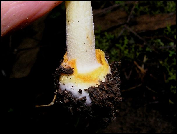
Stem is hollow, 3 to 10cms tall, dry, with hairy scales. Initially white, the stem matures with orange or brown colouration. Whitish to yellowish mycelial threads often appear at the base.

Leratiomyces ceres is reported to be poisonous.

Young Leratiomyces ceres with woolly stems


A bell-shaped cap


Mature stems are sometimes twisted.
Notice pale gills of young specimen.


Gills darken with purplish black spores with maturity.
Notice the white partial veil remnants on rim
and purple-black staining on stem from spores.
(surrounded by tiny Birds Nest Fungi)


Hollow stalk can turn orange with age.


Stem can have a shank buried by wood chips.


Cap ages to brownish orange, gills to dull brown.


My sightings of Leratiomyces ceres

[This will be updated with more sightings]

Hunter Region Botanic Garden, Heatherbrae, NSW - gregarious and clusters in wood chip mulched gardens May 2005, Jul 2010, Aug 2010, Jun 2012.

Barrington Tops National Park, NSW - single fruit body in grassy clearing, Jul 10.

Newcastle University, NSW - wood chip mulched gardens Jun 2012.

A Leratiomyces ceres fruit body infected by a species of the parasitic fungus Spinellus from Family Zygomycete. Sporangiophores are the hair-like reproductive stalks, topped with sporangi (the tiny black dot-like spore-containing receptacles.)

Monday, 19 July 2010

#37 Amanita xanthocephala

This is the first Amanita I've featured on my blog, and it is not a typical Amanita - unlike most Amanitas, it has no ring on the stem.

Amanita xanthocephala (pronunciation: Aman-ee-ta zan-though-seff-allah) is a pretty forest fungus with orange cap and contrasting white gills. It's common name is Vermilion Grisette or Vermilion Amanita.

The orange/yellow/red cap is commonly 3 to 5 cms with a deeper colour towards the centre, and paler similar colour warts. The 'warts' are veil remnants and are generally in the centre of the cap. Initially, the cap is convex, flattening out. The cap margin is radially grooved.

***** ***** *****

The stem is 2 to 6 cms, white to pale yellow, slender, and covered in fine white scales. Although there is no ring on the stem of this Amanita, orange remnants of the volva can be present. The volva (swollen base of stem) is white, but has an out-turned lip bordered in orange or yellow.

Gills are white to pale yellow, close together, and of various lengths. Spore print is white. Amanita xanthocephala is a native fungus found singly or in scattered groups on the ground amongst leaf litter in native forests.

***** ***** *****

This Amanita is mycorrhizal, meaning it forms an association with the host plants' roots where both parties benefit.

A pretty specimen of Amanita xanthocephala
amongst leaf litter and lichen in Werakata National Park


Orange cap with central scales (remains of volva), and striations around margin. Note the orange rim of the volva at ground level.


Although Amanita xanthocephala does not have a stem ring, orange volva remnants can be left on the stem. Also, remnants of the volva can cling to the rim of the cap, as shown in the image below.


Features of the stem and gills. . . . .


. . .and a close-up of the orange out-turned lip of the
volva,
and note the tiny white scales on the stem


A 1cm young Amanita xanthocephala top right of image


A faded aging specimen of Amanita xanthocephala


My sightings of Amanita xanthocephala

[This will be updated with new sightings]

Werakata National Park, Cessnock, NSW - on ground amongst leaf litter and lichen in Eucalypt/Leptospermum/Acacia woodland - Jul 2010, July 2011

Hunter Region Botanic Gardens, Heatherbrae, NSW - amongst leaf litter in garden - Aug 2010, Jun 2011

Wednesday, 23 July 2008

#30 Phallus rubicundus

This pink, orange or red stinkhorn fungus is shaped like a spike or rod, or as the name indicates, is phallic in shape. It grows to about 150mm high and often pops up in lawns, gardens, potplants or mulch.

The matchbox gives an indication of size
of this collapsed Phallus rubicundus


An orange specimen of Phallus rubicundus


This stinkhorn fungus can be distinguished from Mutinus elegans by having separate head holding the brown spore-bearing matter.

As with all stinkhorn fungi, Phallus rubicundus starts out as a gelatinous egg-like structure from which the fruiting body emerges. The fragile hollow stem is perforated, and if sliced horizontally, will be roughly circular. Somewhat sponge-like in texture, it is soft to the touch and will break easily. More information at Mushroom Expert.

The sponge-like texture


The slimy spore-bearing head is separate from the body -
this is an important identifying feature


The interior is hollow and roughly circular.
Notice the stinkhorn "egg" and thin strands of hyphae


The spore-bearing cap has dislodged and slipped down


My sightings of Phallus rubicundus

[This will be updated with new sightings]

Hunter Region Botanic Gardens - Heatherbrae - in mulched gardens, May.

Beresfield Crematorium - in mulched gardens and leaf litter, Dec.

Collapsed and decaying Phallus rubicundus


Tuesday, 22 July 2008

# 29 Pseudocolus fusiformis

Stinkhorns are amongst my favourite fungi. Yes, I know they are considered the "ugly ducklings" of the fungi world, but their offensive reputation is simply a magnet to me. I am always delighted to stumble upon these weird fungi forms, sometimes just having to follow my nose in order to locate them.

I believe this stinkhorn to be Pseudocolus fusiformis, commonly known as Stinky Squid Fungus, although I am not definite about my identification. If any fungi experts out there can confirm or deny my identification, I would be most grateful.

Pseudocolus fusiformis


I have found this stinkhorn fungus on two occasions, both in mulch at the Hunter Wetlands at Shortland, Newcastle. It is the foulest smelling stinkhorn species I have encountered, resembling the stench of a blocked or unmaintained septic sewer system. The odour emanated for quite a distance.

Arms of Pseudocolus fusiformis emerging from leaf-litter


When leaf-litter is scraped away the "eggs" can be seen


Like all stinkhorn fungi, Pseudocolus fusiformis starts out as a gelatinous egg-like form buried or semi-buried in the substrate. As it ages, the fruit body emerges from the "egg". Up to 4 arms are joined to a single "stem" and are joined at the apex.

The green/brown or brown slimy spore-bearing gleba is present on the inside surface of the arms. The foul odour attracts flies which distribute the spores.

There is more information on Pseudocolus fusiformis at Mushroom Expert.


Notice the gelatinous substance in the ruptured "egg"


My sightings of Pseudocolus fusiformis

[This will be updated with new sightings]

Hunter Wetlands (Shortland, Newcastle) - in mulch or leaf-litter - Jun, Dec.


Arms are joined at apex and gleba is presented
on the inside surface of the arms

Thursday, 15 November 2007

#28 Anthracophyllum archeri


Anthracophyllum archeri, commonly known as Orange Fan, is from family Marasmiaceae, and grows on dead wood in native forests. Pronunciation is Anthrack-oh-fill-um archer-eye. It is a common fungus and can fruit at any time of year.

Caps are shell-shaped or fan-shaped, up to 35mm and are attached laterally to wood, occasionally with a very short stem, but my local observations have noted the absence of a stem. The upper surface of the cap is smooth and radially ridged, tan to light orange.

Smooth ridged upper surface of Orange Fan Fungi


Gills are similar in colour but often darker, widely spaced and fan out in various lengths from the point of attachment. Fungi have a smooth leathery texture.

Orange Fan Fungi are usually seen in large numbers and can be spaced along a stick or log, or crowded forming overlapping clusters. The structure of the fan shape with widely spaced fold-like gills make Anthracophyllum archeri easily distinguished from other fan-like or shell-like fungi.


Orange Fan Fungi occur as many single fruiting bodies.....


.....or crowded overlapping clusters.


My sightings of Anthracophyllum archeri

[This will be updated with new sightings]

Barrington Tops National Park NSW - on dead twigs and logs in rainforest - Mar, Apr, May, Nov.

Hunter Region Botanic Gardens - Heatherbrae NSW - on dead twigs in open eucalypt forest - Nov.

Friday, 21 September 2007

#25 Stereum ostrea


Stereum ostrea, Golden Curtain Crust, is a leathery shelf fungus belonging to family Stereaceae. Pronunciation Steer-ee-um Austria. This common fungus often makes a spectacular, long-lasting, massed display. It is found in many parts of the world, and internationally it is known as 'False Turkey Tail'.

It is found on dead wood (fallen or standing) in wet native forests, and its Australian distribution includes the eastern coastal and mountainous areas from north Queensland, through NSW, Victoria and Tasmania.

Attractive concentric zones and soft leathery texture


Fruiting bodies are broadly attached to the substrate, either singly or often fused laterally forming overlapping rows and commonly colonising whole logs, remaining fresh-looking for months. The thin shelf, fan or funnel-shaped fruit-bodies are only about 2mm thick with concentric (circular) bands or zones of yellow, orange and many shades of brown, usually with a pale wavy margin. Brackets can be up to 100mm, or more, in diameter.

The upper suface of fruit-bodies feels ridged but smooth, and 'velvety' with very short hairs. Stereum ostrea has a smooth underside which distinguishes it from the somewhat similar Trametes versicolor which has pores on the underside. Spore print is white. I have not successfully obtained a spore print of Stereum ostrea.

Fused fruit bodies form overlapping rows of 'shelves'


Smooth but lumpy lower surface of Stereum ostrea can be white, yellow, orange or gold. Illustrated in the image, fruit-bodies can be adhered to the underside of logs with only the fertile underside visible. All fungi surfaces here are the lower surface.


The sun shines through the thin leathery fungi 'fans' and highlights the orange circular bands. Notice the narrow band of cream lining the margin, which is a feature of Stereum ostrea.


My sightings of Stereum ostrea

[This will be updated with new sightings]

Barrington Tops National Park, NSW - on felled logs in damp forest - Jul, Sep.

A massed display of Stereum ostrea

Tuesday, 22 May 2007

#15 Lactarius deliciosus


Lactarius deliciosus, Saffron Milk Cap, belongs to the family Russulaceae. Lactarius species exude latex (milky fluid) when cut. When cut, the latex stains the brittle flesh bright orange. This is an introduced species that forms a mycorrizal association (symbiotic fungal dependency) with introduced pines.

Orange gills and blotched stem of young specimen


The cap is convex (rounded or domed) at first, with a central depression, becoming broadly funnel-shaped. Surface texture is smooh and dry, but is often viscid (slimy) when wet. Spore print is pale yellow, and cap can reach 160mm diameter.

When young, the cap is carrot-orange or sometimes a dull apricot colour, and can have concentric (circular) zones of colour. Caps and stalks become a dull greenish colour when aged or bruised.

A cut fungus displays the cut surface stained bright orange

Gills are orange at first, bruising greenish with age, decurrent (running down the stem), and various lengths. The stem is stout and up to 70mm in height, blotched orange when young, often becoming greenish with age.

This species is reported to be edible with the correct preparation, but I can not vouch for its palatability. I am unsure as to the distribution of Lactarius deliciosus.

Illustrating the sometimes apricot cap and circles of colour

My sightings of Lactarius deliciosus

[This will be updated with more sightings]

Hanging Rock, New England region of NSW - in and near pine plantations: Mar, Apr, May.

An aged specimen with a matchbox for size comparison

Tuesday, 15 May 2007

#11 Dermocybe splendida


Dermocybe splendida has the common name of Splendid Red Skinhead, and is from family Cortinariaceae. Pronunciation is Dermo-sigh-be splendid-da.


The Cortinariaceae group of fungi are typically recognised by the presence of a 'cortina'. The cortina is a veil of silky or cobwebby fibrous threads which protects the gills of a young mushroom. The veil collapses as the cap opens in most cases, often leaving hairs on the stem which then stain brown from the spores.


Young Dermocybe splendida with veil covering gills


In the image below, the fibrous veil has been stretched as the cap expands. And in the image below this, the cobwebby strands have almost completely separated from the cap.

The cortina (fibrous veil) tearing as the cap expands


And the veil is almost completely separated from cap


This attractive fungus grows in soil in Eucalyptus and Leptospermum (tea-tree) forests in eastern NSW, Victoria, Tasmania, south-eastern SA, and south-western WA.

Orange-red caps are initially convex (domed), expanding up to 90mm into a broadly umbonate red-brown cap. An 'umbo' is a dome-like swelling in the centre of the cap. A slight umbo can be seen in the fungus on the right of the first photograph. The cap is dry and smooth. This fungus can be found singly, or in small or large groups.

The stem can reach 130mm in height, tapering upwards, and can be bulbous at the base. The tapering of the stalk is evident in the following image, and a bulbous base can be seen in the previous picture.

Two distinguishing features are the bright paprika-red gills, and the yellowish base of the brown-red stem and yellow mycelium. The mycelium is a mass of underground filaments (hyphae) which are often visible at the base of the fungus. The yellow mycelium is noticable in the foreground of the next image.

A dusting of rust-brown spores can be seen on caps in the following picture. This is a common occurence amongst Cortinariaceae as spores drop from overhanging fungi. The red gills stain brown as the cap ages.

Young and mature Splendid Red Skinheads


My sightings of Dermocybe splendida

[This will be updated with more sightings]

Hanging Rock, NSW - Eucalyptus woodland with tussock grass - Jun.

Barrington Tops National Park, NSW - Rainforest, in leaf litter - Apr.

New England National Park, NSW - Eucalyptus woodland with tussock grass - May.